
X-ray Applications
X-rays have a wide range of applications in various fields due to their ability to penetrate matter and provide valuable information about the internal structure of objects. Some common applications of X-rays include [6]:

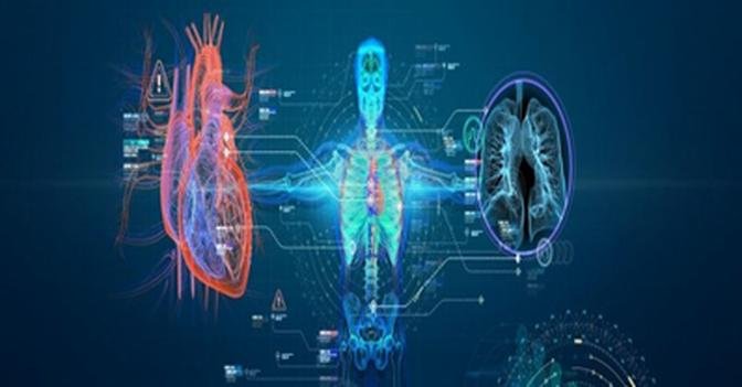
- Medical Imaging[7]:
Radiography: X-rays are extensively used in medical radiography to visualize the internal structures of the human body. This includes X-ray examinations of bones, tissues, and organs, helping diagnose fractures, infections, tumors, and other medical conditions.
- Computed Tomography (CT): CT scans use X-rays to create detailed cross-sectional images of the body, allowing for the diagnosis and monitoring of a wide range of medical conditions.
- Fluoroscopy: This real-time imaging technique uses X-rays to visualize the movement of internal structures, often used during procedures like angiography and barium swallow studies.
- Mammography: an x-ray imaging method used to examine the breast for the early detection of cancer and other breast diseases. It is used as both a diagnostic and screening tool.
- Dentistry: X-rays are used in dental radiography to detect dental issues like cavities, gum disease, and impacted teeth.
- Airport Security: X-ray scanners are used at airports to screen luggage and carry-on items for concealed objects or prohibited items.
- Non-Destructive Testing (NDT):
- Industrial Radiography: X-rays are used to inspect welds, pipelines, and other industrial components for defects without damaging the material.
- X-ray Fluorescence (XRF): XRF is used to determine the elemental composition of materials, helpful in fields like metallurgy, archaeology, and environmental science.
- Materials Science: X-ray diffraction and scattering techniques are used to study the atomic and molecular structures of materials, aiding in the development of new materials and pharmaceuticals.
- Security Scanning: X-ray scanners are employed in security screening at entrances to detect concealed weapons or contraband.
- Crystallography: X-ray crystallography is used to determine the three-dimensional structures of molecules, including proteins and other biological macromolecules.
- Archaeology and Art Conservation: X-ray imaging is used to examine the internal structure of archaeological artifacts and artworks without damaging them.
- Food Inspection: X-rays are used to inspect packaged food products for contaminants, foreign objects, and quality control.
- Oil and Gas Industry: X-rays are used for imaging subsurface structures in the exploration and production of oil and gas reservoirs.
- Astronomy: X-rays from celestial objects provide insights into high-energy astrophysical processes, such as the study of black holes, neutron stars, and galaxy clusters.
- Radiation therapy: also known as radiotherapy, is the use of X-rays for therapeutic purposes and is widely used in the treatment of cancer. It requires higher radiation doses than X-ray imaging. Radiation therapy can treat various forms of cancer, including skin cancer and internal tumors such as those in the brain, lungs, prostate, and breast, among others. However, radiation therapy itself can cause significant harm to the human body and has the potential to induce cancer.
X-ray Vision and Perspective Abilities
After Wilhelm Röntgen's discovery of X-rays, he quickly recognized their ability to provide perspective, similar to the visual capabilities of human "X-ray vision." While X-rays are invisible to the naked eye, Röntgen and other researchers found that some individuals appeared to have X-ray vision. Röntgen himself once noticed a blue light coming from an X-ray tube that seemed to originate from his own eyes. However, at the time, he dismissed this observation, believing it to be an anomaly associated with a specific type of X-ray tube he was using. Later, Röntgen realized that the X-ray tube responsible for this phenomenon was his only high-powered X-ray tube, and that X-rays became visible when the power was sufficiently high, making the experiment easy to replicate. Nevertheless, this phenomenon was only reported by a few individuals, suggesting that most people could not see X-rays.
Due to the destruction of all experimental records after Röntgen's death and the significant harm X-rays can cause to the human body, such experiments have long been prohibited in the academic community. However, some individuals do seem to possess X-ray vision-like abilities even without direct exposure to X-rays. For example, a Russian woman named Natalya Demkina (1987-) is believed to have X-ray vision. Demkina participated in multiple experiments that indicated she had X-ray-like perspective, enabling her to directly observe the skeletal and visceral organs inside the human body and detect any abnormalities. Her diagnoses corresponded with those obtained from X-ray imaging in hospitals. In 2004, the Discovery Channel produced a documentary titled "The Girl with X-ray Vision," where researchers presented Demkina with six X-ray diagnostic reports and asked her to observe six afflicted volunteers and one asymptomatic volunteer, and determine which diagnostic report matched each volunteer or if the volunteer was not afflicted. Demkina successfully matched four out of seven individuals, including the asymptomatic volunteer. However, researchers believed Demkina needed to match at least five individuals to demonstrate statistical significance, and thus, they were skeptical of the authenticity of her X-ray vision.
However, Nobel Prize-winning physicist Brian Josephson (1940-) from the University of Cambridge argued that the statistical methods used by the researchers were flawed. He suggested that the probability of Demkina successfully matching four out of seven individuals by luck alone was only 2%, and therefore, the experiment actually confirmed the existence of X-ray vision. In fact, there are historical records of ancient Chinese medical practitioners such as Bian Que and Hua Tuo possessing perspective abilities, even though the concept of X-rays did not exist at that time. These individuals could see things with their naked eyes that were normally hidden, suggesting that X-ray vision-like abilities may be a subset of the broader concept of enhanced vision.
Modern science also provides other examples of X-rays being visible. For instance, if the intensity of X-rays is high enough, it can ionize molecules in the air, causing an ionization phenomenon that is directly visible to the naked eye. Facilities like the European Synchrotron Radiation Facility can emit high-power X-ray beams capable of causing ionization phenomena that are visible to the human eye.
X-ray Adverse Effects
X-rays are a valuable tool in medicine and industry, but like any form of ionizing radiation, they can have adverse effects on human health when exposure is excessive or not properly managed. Adverse effects of X-ray exposure can range from short-term, acute effects to long-term, chronic effects. Here are some of the adverse effects associated with X-ray exposure[8-11]:
1. Tissue Damage: High doses of X-rays can cause immediate tissue damage. This can manifest as radiation burns or radiation sickness, which may include symptoms such as redness, blistering, nausea, vomiting, and fever. Acute radiation syndrome (ARS) is a severe form of radiation sickness that can be life-threatening.
2. Cancer Risk: Prolonged or repeated exposure to X-rays can increase the risk of cancer. Ionizing radiation, including X-rays, is a known carcinogen. The risk is particularly elevated for organs and tissues that are sensitive to radiation, such as the thyroid, breast, and lungs.
3. Genetic Damage: X-rays have the potential to cause genetic mutations and other cellular changes by damaging DNA. While the risk of genetic mutations is generally low for diagnostic X-ray procedures, it is a concern in high-dose or repeated exposures.
4. Radiation-Induced Cataracts: High-dose or chronic exposure to X-rays can lead to the development of radiation-induced cataracts, which can impair vision. The lens of the eye is particularly sensitive to ionizing radiation.
5. Radiation-Induced Skin Conditions: Overexposure to X-rays can lead to radiation dermatitis, characterized by skin redness, itching, and potential skin damage.
6. Fetal and Developmental Effects: Pregnant women are particularly vulnerable to the adverse effects of X-rays. Ionizing radiation can harm the developing fetus, leading to birth defects, developmental issues, or an increased risk of childhood cancer.
7. Long-Term Health Risks: Cumulative exposure to X-rays over time can contribute to long-term health risks, including an elevated risk of cancer and other radiation-related diseases.
There is ongoing debate about the concept of radiation hormesis, which suggests that low-dose radiation exposure may have health benefits or stimulate protective responses in the body. This is a controversial and not widely accepted idea, and current radiation protection guidelines are based on the linear no-threshold (LNT) model, which assumes that any level of ionizing radiation carries some risk.
It's important to emphasize that the adverse effects of X-ray exposure are generally associated with high doses or prolonged and repeated exposures. In medical settings, X-ray procedures are carefully controlled to minimize radiation exposure to patients, and the benefits of obtaining diagnostic information often outweigh the associated risks.
Healthcare providers and radiologic technologists follow strict protocols and guidelines to ensure that X-ray exams are conducted safely and with the least possible radiation dose. Patients should discuss any concerns about X-ray exposure with their healthcare providers, especially if they are pregnant or have a history of radiation exposure. Proper safety measures, adherence to radiation protection principles, and minimizing unnecessary X-ray exams are essential in managing and mitigating X-ray-related risks.
X-ray Measurement
Measuring X-rays involves using various instruments and detectors that can quantify the radiation's intensity, dose, or energy. The choice of measurement method depends on the specific application, whether it's ensuring patient safety in medical settings, quality control in industrial testing, or conducting scientific research [12].
Here are some common methods for measuring X-rays:
1. X-ray Dosimeters:
- X-ray dosimeters are devices specifically designed to measure and record the dose of X-ray radiation received by an individual or object. These dosimeters are essential for ensuring radiation safety in medical and industrial settings.
- Types of X-ray dosimeters include film badges, thermoluminescent dosimeters (TLDs), optically stimulated luminescence dosimeters (OSLDs), and electronic personal dosimeters (EPDs). These devices are worn by individuals to monitor their radiation exposure.
2. Geiger-Muller (GM) Counters:
- GM counters are versatile radiation detectors that can measure various types of ionizing radiation, including X-rays. They work by detecting the ionization caused by X-ray photons in a gas-filled chamber.
- GM counters produce an audible clicking sound or a visual display of counts per unit of time, allowing users to assess the level of X-ray radiation in the vicinity.
3. Ionization Chambers:
- Ionization chambers are used to measure X-ray exposure and dose. They consist of a gas-filled chamber where X-rays ionize the gas molecules, creating an electric current that is proportional to the radiation dose.
- These chambers are often used in medical physics and radiation therapy to ensure precise dose delivery in treatments.
4. Solid-State Detectors:
- Solid-state detectors, such as silicon or germanium detectors, are semiconductor devices that directly convert X-ray energy into electrical signals. These detectors are widely used in research and scientific experiments.
- They offer high sensitivity, energy resolution, and precision for measuring X-ray energies and intensities.
5. Scintillation Detectors:
- Scintillation detectors use scintillating materials that emit visible or ultraviolet light when exposed to X-rays. Photodetectors then convert the emitted light into an electrical signal.
- These detectors are commonly used in medical imaging, nuclear medicine, and high-energy physics experiments.
References
1. Glasser, O., Röntgens vorläufige Mitteilung „Über eine neue Art von Strahlen“, in Wilhelm Conrad Röntgen und Die Geschichte der Röntgenstrahlen, O. Glasser, Editor. 1931, Springer Vienna: Vienna. p. 12-21.
2. Price, R. and W. Morgan, Electrical Experiments Made in Order to Ascertain the Non-Conducting Power of a Perfect Vacuum, &c. By Mr. William Morgan; Communicated by the Rev. Richard Price, LL.D. F.R.S. Philosophical Transactions of the Royal Society of London, 1785. 75: p. 272-278.
3. Anderson, J.G., William Morgan and X-rays. Transactions of the Faculty of Actuaries, 1945. 17: p. 219-221.
4. Stöhr, J., Introduction and Overview, in The Nature of X-Rays and Their Interactions with Matter, J. Stöhr, Editor. 2023, Springer International Publishing: Cham. p. 1-58.
5. Stöhr, J., The Nature of X-Rays and Their Interactions with Matter 2023: Springer; 1st ed. 2023 edition (June 8, 2023).
6. Brown, J.G., X-Rays and Their Applications 2011: Springer New York, NY.
7. Mould, R.F., A Century of X-Rays and Radioactivity in Medicine 2019: CRC Press
8. Toppenberg, K.S., D.A. Hill, and D.P. Miller, Safety of radiographic imaging during pregnancy. Am Fam Physician, 1999. 59(7): p. 1813-8, 1820.
9. Berrington de González, A. and S. Darby, Risk of cancer from diagnostic X-rays: estimates for the UK and 14 other countries. Lancet, 2004. 363(9406): p. 345-51.
10. Chauhan, V. and R.C. Wilkins, A comprehensive review of the literature on the biological effects from dental X-ray exposures. International Journal of Radiation Biology, 2019. 95(2): p. 107-119.
11. Wagner, L.K., P.J. Eifel, and R.A. Geise, Potential Biological Effects Following High X-ray Dose Interventional Procedures. Journal of Vascular and Interventional Radiology, 1994. 5(1): p. 71-84.
12. Taylor, L.S., The Precise Measurement of X-Ray Dosage. Radiology, 1930. 14(4): p. 372-384.